
LIZFRANC INJURIES

Diagnosing and Treating Lizfranc Injuries
ACE Physical Therapy and Sports Medicine Institute
Tips for Lizfranc Injuries.
- Lizfranc joint is named after Jacques Lizfranc, a field surgeon in Napoleon’s army who performed an amputation of a soldier’s foot in this region.
- Lizfranc fracture dislocations account for approximately 1% of all foot injuries.
- Many high level athletes that suffer a Lizfranc fracture dislocation never return to their pre-injury status.
- Un-diagnosed lizfranc injuries can lead to the pre-mature onset of arthritis in the foot.
- Follow the protocols set by your doctor and physical therapist and have the best chance to return to your pre-injury status.
The simple task of walking across the room can become extremely difficult if you are suffering from Lizfranc injury. Unfortunately, this uncommon injury is often misdiagnosed as a sprain or even undiagnosed. This can lead to chronic pain and even loss of function. The good news is that Lizfranc is treatable. A proper diagnosis and treatment increases recovery rate and helps prevent long-term problems.
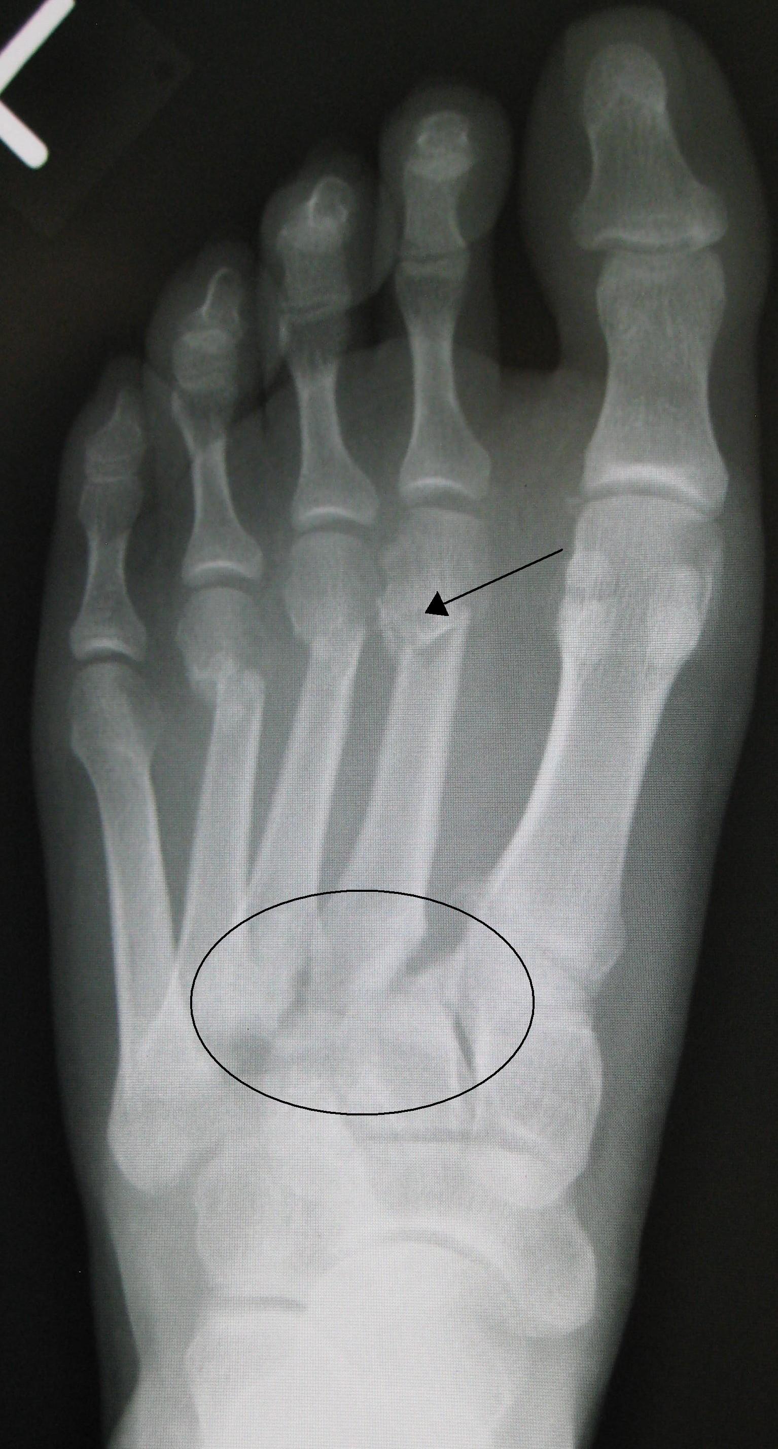
The anatomy of the human foot is labeled into three distinct parts, the hind (rear), mid and forefoot. The Lizfranc joint or complex is comprised of the medial aspect of the mid-foot or mid-tarsal joint. The joint is made up of the 1st and 2nd metatarsal and 1st and 2ndcuneiform bones. The proximal end of the 2ndmetatarsal or base “wedges” itself into the “keystone” or 2nd cuneiform bone. This bony alignment leads to a very stable juncture that is capable of transferring the forces from the rear or hind foot to the forefoot during gait. The joint is held together on the bottom or plantar surface with a thick ligament (leather-like in nature) that is capable of withstanding hundreds of pounds of force without being disrupted. The superior aspect of the joint is not well stabilized and is the “weak” part of the joint. When an injury occurs, there are varying degrees of damage to the structures of the joint.
 The most common type of injury to the Lizfranc joint is a common, grade I sprain. It involves the mild damage to the ligaments. There is no displacement of the bones and no laxity to the joint. Pain, bruising, swelling and difficulty bearing weight are natural symptoms that are found with this type of injury. More severe injuries include grade II sprains which include some laxity and displacement of the bones. The most severe injury is a grade III sprain, which involves complete rupture of the ligaments and dislocation of the bones.
The causes of Lizfranc injuries are numerous such as high energy or low energy traumatic events to the joint. High-energy traumas may include auto accident or falling from a high location and landing on the foot. Low energy trauma occurs when the foot is twisted or the foot is “planted” and stationary and the leg is twisted, producing a torque through the joint. This typical happens on the athletic fields or during an accidental slip and fall.
Diagnosing these injuries is difficult. A normal x-ray does not usually reveal any damage to the joint because it is taken in a non-weight bearing position. If the x-ray is performed in the weight bearing position it often times reveals a larger gap or space between the cuneiforms and this is an indication that the joint has sustained some damage and laxity most likely is present. More advanced testing, MRI and CT scans are not required to diagnose a Lizfranc injury, but some doctors rely on these tests if there are question about the diagnosis or if surgery is required to fix the damaged joint.
Treatment of these injuries can be controversial. Some healthcare professionals differ in opinion on the treatment of a grade II injury because there is not gross instability that is found in a grade III injury, but there is much more excess motion in the joint when compared to the bones of a grade I injury. The difference in op bones with plates and screws or to immobilize the joint with a cast for a prolonged period of time. All treatments options will require the patient function in a non-weight bearing status for several days to weeks.
Physical Therapy treatment can begin immediately. The patient’s treatment protocol must respect and adhere to the strict non-weight bearing status. The patient should be taught numerous “open-chain” exercises to help maintain the muscle strength in the involved lower extremity. The treatment protocol must incorporate core exercises. While the patient is non-weight bearing status, there is a danger in weakening the muscle structure of the entire lower extremity: don’t use it and lose it. When the patient is “cleared” to weight bear, different, more aggressive exercises will have to be prescribed to return the involved lower extremity back to its pre-injury status.
The formal physical therapy program will include the use of manual techniques and modalities to reduce swelling and edema when the foot is exposed. The entire foot/ankle complex will be swollen and the motion will be limited. The non-involved joints will need attention to regain their motion and function and this can be accomplished while the injured Lizfranc joint heals. While the rehabilitation program will be slow going initially, most patients should be able to return to pre-injury status and activity level.
Lizfranc injuries are not common. When they do occur, they are often times missed in the evaluation process. Proper and quick diagnosis of the injury by an astute healthcare professional can prevent long-term chronic problems in the involved foot and make a return to pre-injury status possible.
Read more articles on our main website blog at: ACE-pt.org/blog
Vist our main website at www.ACE-pt.org